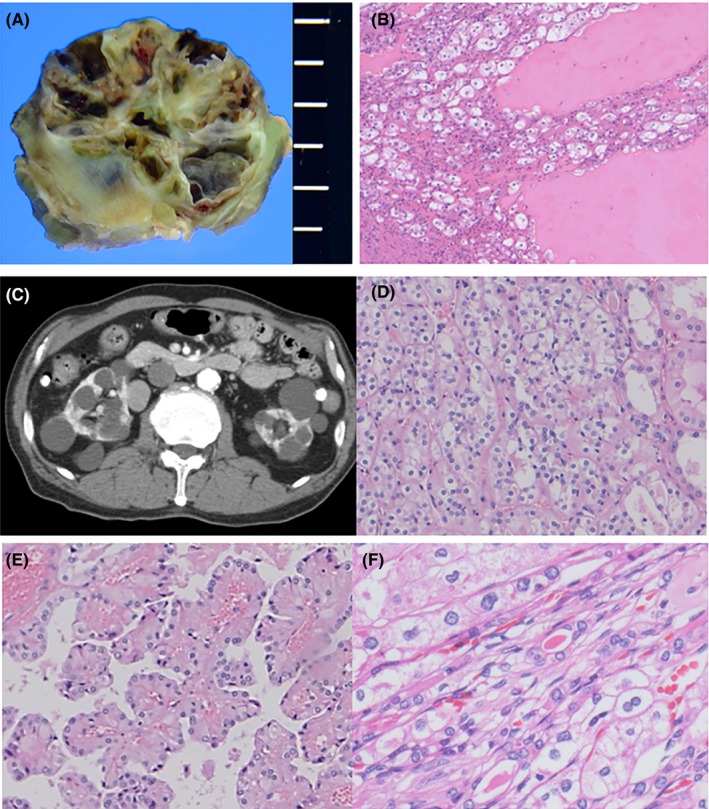
Figure 2.

Macro‐ and microscopic features of cystic tumors. A, Gross section of a cystic tumor in a 61‐year‐old man. B, Microscopic features of (A). Tumor cells composed of oncocytic and clear cells are present along the cyst walls. The cysts are filled with eosinophilic exudate. C, Abdominal computed tomography scan of a 65‐year‐old man with chronic renal failure and dialysis. Polycystic lesions are present in both kidneys. D‐F, Microscopic features of the resected kidneys in (C). Tumors are composed of hybrid (D), papillary adenomatous (E) and chromophobe‐like morphology with raisinoid nuclei and/or binucleation (F)
