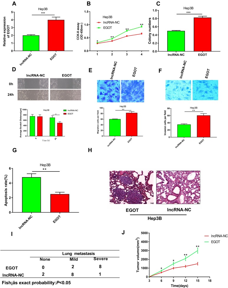
Figure 3.
Overexpression of EGOT promoted the malignant phenotypes of HCC cell line Hep3B. (A) qRT-PCR was used to verify the efficiency of overexpression of EGOT in Hep3B cells. (B) CCK-8 method was used to detect the proliferation of Hep3B cells after EGOT overexpression. (C) Colony formation assay was conducted to evaluate the ability of colony formation of Hep3B cells after EGOT overexpression. (D) Wound-healing assay was used to examine the motility of Hep3B cells after EGOT overexpression. (E, F) Transwell assays were used to detect the migration and invasion of Hep3B cells after EGOT overexpression, respectively. (G) The apoptosis of Hep3B cells after EGOT overexpression was analyzed by flow cytometry. (H, I) Incidence and severity of lung metastasis in mice pulmonary metastasis model with EGOT overexpressed Hep3B cell and control cell. (J) Tumor volume in nude mice xenograft model with EGOT overexpressed Hep3B cell and control cell. *P<0.05, **P<0.01, ***P<0.001.

