Figure 4.
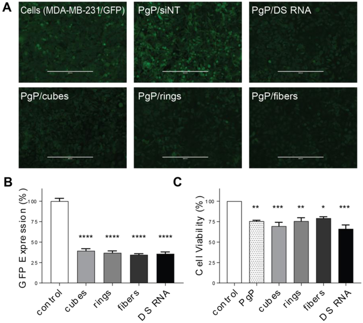
Specific gene silencing with PgP/NANP(GFP) polyplexes and cell viability assays tested against GFP expressing breast cancer cells (MDA-MB-231/GFP). Fluorescent microscopy (A) shows representative images of GFP knockdown by PgP/NANP(GFP) polyplexes. (B) GFP knockdown efficiency by cubes (PgP/cubes(GFP)), rings (PgP/rings(GFP)), and fibers (PgP/fibers(GFP)) are compared to free DS RNAs(GFP) and negative control (siNT), PgP only (0.1 mg/mL) used as an additional control. (N=3, **** denotes statistically significant vs. untreated cells with p<0.0005, # denotes statistical significance vs. cubes with p<0.05). (C) Cell viability by PgP/NANP(GFP) polyplexes (N=3, * denotes statistical significance with p<0.05, ** with p<0.005, *** with p<0.005).

