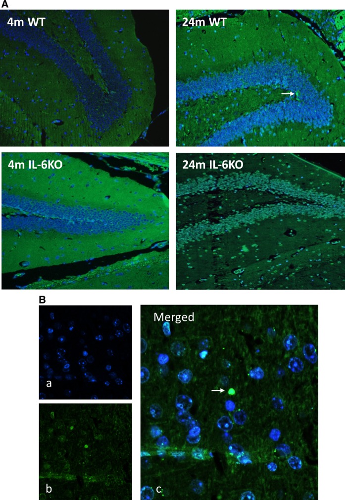
Fig. 3.
a Representative microphotographs of Hoechst 33258 and TUNEL staining of hippocampal dentate gyrus form 4- and 24-month-old IL-6-deficient (IL-6KO) and wild type control (WT) mice revealed single apoptotic cells in hippocampus of aged mice of both genotypes, (magnification × 200). b (a) Fluorescent Hoechst 33258 (blue) nuclear staining, (b) TUNEL staining (green) indicating apoptotic nucleus, (c) Merged images of Hoechst 33258 (blue) and TUNEL (green) staining, (magnification × 400). White arrow indicates apoptotic cell

