Figure 3.
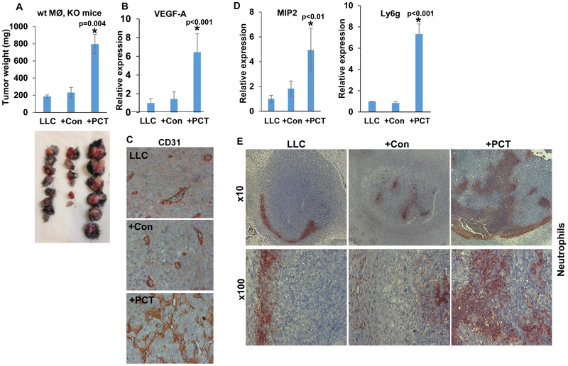
PCT-treated macrophages promote tumor growth and vascularity. A. Tumor growth. LLC cells (4×10⁵) were inoculated subcutaneously in Hpa-KO female mice (8-10 weeks old) without (LLC; n=8) or with an equal number of untreated (+Con; n=6) or PCT-treated (+PCT; n=6) macrophages. At termination on day 21, tumors were excised, weighed (upper panel) and photographed (lower panel). *p=0.004 +PCT vs LLC/+Con. Total RNA was extracted from a portion of the tumors and subjected to qPCR applying VEGF-A specific primers (B; p<0.01 +PCT vs. LLC/+Con). The rest of the tumors were fixed in formalin, embedded in paraffin and five-micron sections were subjected to immunostaining applying anti-CD31 antibody (C). Note increased vascularity of LLC tumors inoculated with PCT-treated macrophages. Original magnifications: x100. Tumor samples were similarly subjected to qPCR applying primers specific for MIP2 (D, left) and Ly6g (a marker for neutrophils; D, right). p<0.01 +PCT vs. LLC). E. Tumor sections were subjected to immunostaining applying anti-Ly6g antibody. Note the recruitment of neutrophils to LLC tumors implanted together with PCT-treated macrophages. Original magnifications: upper panels x10, lower panels: x100.

