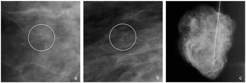
Figure 2. Case example of patient with no pMRI.
(a-c) Patient with biopsy proven intermediate grade DCIS with a 10 mm group of linear calcifications (circles) seen on cropped CC (a) and MLO (b) mammographic views. No pMRI was obtained and a wire-guided lumpectomy was performed with the surgical specimen (c) showing the targeted clip and wire. Pathology revealed DCIS spanning at least 22 mm with positive margins. The patient then underwent a skin-sparing mastectomy.

