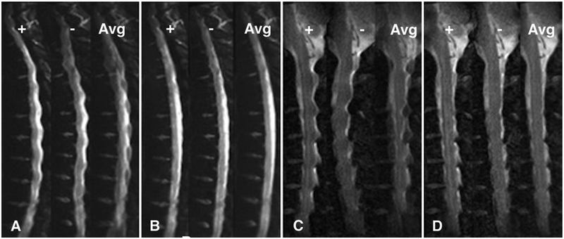
FIG. 3.
Thoracic and cervical b = 0 images demonstrating positive distortion (+), negative distortion (−), and the average of both directions of distortion (Avg). A and C: b = 0 images where no distortion correction has been applied. B and D: The same images after performing distortion correction with the displacement field calculated from the positive (+) and negative (−) images.

