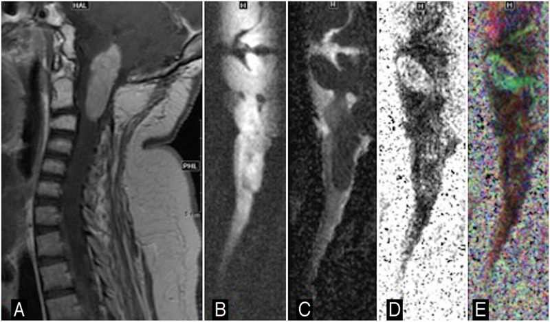
FIG. 6.
Images obtained in a 6-year-old girl with a brainstem pilocytic astrocytoma. Sagittal contrast-enhanced T1-weighted image (A), sagittal diffusion-weighted image (B), sagittal ADC (C), sagittal FA (D), and sagittal colored FA map (E). Note the position of the fourth ventricle relative to mass.

