
Figure 4. Quantitative analysis of particle distribution relative to total tumor area and BBB breakdown demonstrates increased particle diffusion with cRGD targeting at 96 hours post-treatment.
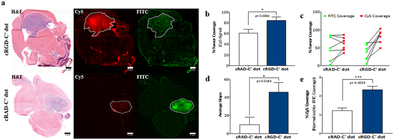
RCAS-tumor bearing mice were treated with cRGD- or cRAD-C’ dots and sacrificed 96 hours later. FITC-Dextran was administered 3 hours prior to sacrifice. Fluorescence analysis of FITC and Cy5 signal on frozen sections was performed and total tumor areas were measured using ROI analysis of H&E staining. a) Representative images of mGBM brain sections of mice treated with cRGD- (top row) versus cRAD-C’ dots (bottom row) 96 hours post-treatment. b) Comparison of cRAD-C’ dot and cRGD-C’ dot tumor coverage as measured by Cy5 fluorescent signal (Student’s T-test, * p = 0.0304). c) Tumor coverage of administered FITC-Dextran and cRAD- or cRGD-C’ dots across all experimental animals. d) Average slope comparison of cRAD- and cRGD-C’ dot treated animals depicted in Figure 4c (Student’s T-test, * p = 0.0184). e) Cy5 percent tumor coverage normalized to average FITC signal in both cRAD- and cRGD-C’ dot treated tumors (Student’s t test, *** p = 0.0003). (Scale bars = 1 mm)