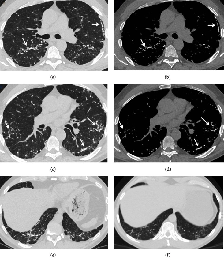
Figure 1.
Axial chest CT images using lung (a), (c) and bone (b), (d) windows show bilateral branching dense nodular opacities (arrows) with mild associated reticulation. Some of the nodule are high in attenuation and almost iso-dense to ribs on bone windows. Axial images using lung widow (e), (f) at the level of lung bases were obtained 5 years apart and show evidence of progression.

