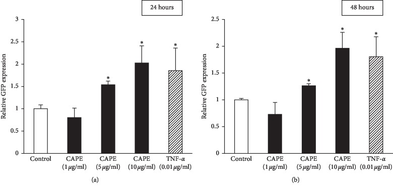
Figure 8.
NF-κB signal pathway in CAPE-stimulated KN-3 cells. A GFP reporter construct with transcriptional response element for NF-κB was transiently transfected into KN-3 cells and subjected to stimulation with CAPE (1, 5, or 10 μg/ml) for 24 (a) or 48 hours (b). Treatment with TNF-α (0.01 μg/ml) was used as a positive control. The expression level of GFP was measured by using a fluorescence microplate reader. Values represent the means ± SDs from representative of two independent experiments, and each experiment was performed in quadruplicate. Asterisks indicate significant differences versus the control (medium alone) (∗p < 0.05 vs. control).

