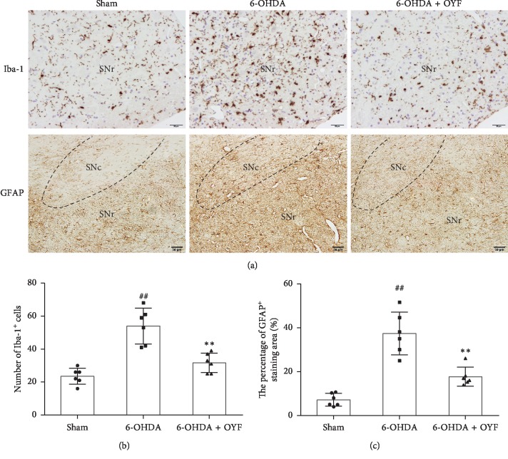
Figure 4.
Effects of OYF on Iba-1 and GFAP expressions in the SN of 6-OHDA-induced PD rats. (a) Representative images of Iba-1 and GFAP-positive cells in the ipsilateral SN, scale bars = 20 μm (Iba-1) and 50 μm (GFAP). (b) The number of Iba-1-positive cells in the ipsilateral SN. (c) The percentage of GFAP-positive staining area in the ipsilateral SN. Values are expressed as mean ± SD. n = 6 per group. ##P < 0.01 vs. the sham group, and ∗∗P < 0.01 vs. the 6-OHDA group. SNc, SN compacta; SNr, SN reticulate.

