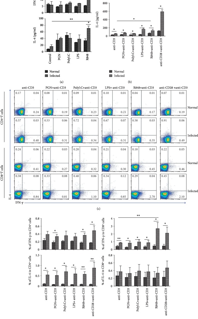
Figure 3.
Role of TLR agonists in inducing IFN-γ and IL-4. Single mononuclear MLN cell suspensions of normal and infected mouse were prepared and cultured in vitro with PGN, PolyI:C, LPS, and R848, with or without anti-CD3 Ab. 72 h later, the concentration of IFN-γ (a) and IL-4 (b) in the supernatants of cultured cells was detected by ELISA. The MLN lymphocytes isolated from normal and infected mice were stimulated by PGN, Poly I:C, LPS, and R848, with anti-CD3 Ab. The expression of INF-γ and IL-4 on CD4+ or CD8+ T cells in normal and infected mouse was detected by flow cytometry. (c) The numbers represent the expression of cells in each subset. The average percentages of INF-γ and IL-4 in CD4+ (d) or CD8+ (e) T cells were calculated from the FACS analysis. Three independent experiments (5–6 mice per group) were performed, and one representative result is shown. ∗P < 0.05, ∗∗P < 0.01, nsP > 0.05.

