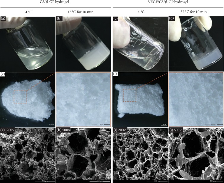
Figure 1.
The process of gelation and the microstructure of hydrogels. Photographs of CS/β-GP gel before and after gelation (a, b). Photographs of VEGF/CS/β-GP gel before and after gelation (c, d). Photographs of CS/β-GP and VEGF/CS/β-GP hydrogels after lyophilization (e, f). SEM images of CS/β-GP hydrogel in 200x and 500x (g, h). SEM images of VEGF/CS/β-GP hydrogel in 200x and 500x (i, j).

