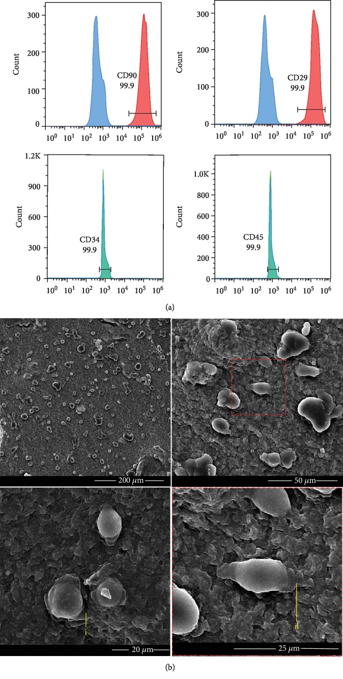
Figure 2.
Cell surface markers on DPSCs and the morphology of DPSCs cultured on the hydrogel. Flow cytometric analysis was used to test the surface markers of DPSCs. DPSCs were positive for CD29 and CD90, and negative for CD34 and CD45 (a). Morphology of DPSCs cultured on the surface of CS/β-GP hydrogel after 24 h (b). DPSCs embedded their cellular synapses into the pore canal (i, ii).

