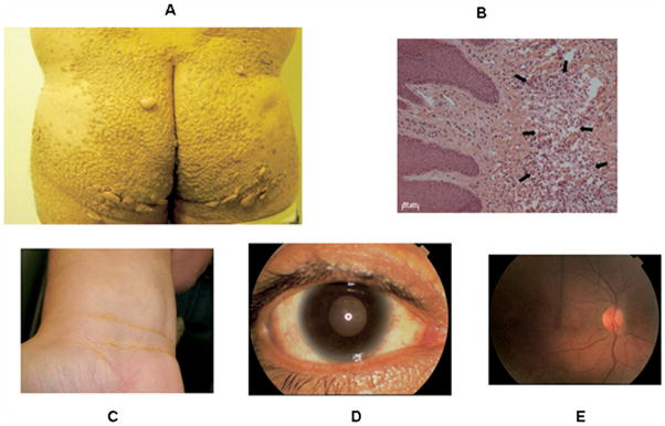
Figure 1.
(A) Tubero-eruptive xanthomas on the buttocks of a patient with homozygous familial apolipoprotein A-I deficiency as described in Matsunaga and colleagues.28 (B) Light microscopy of a biopsy of one of these xanthomas documenting lipid-laden macrophages. (C) Planar xanthomas in the skin creases of the wrist. (D) Photograph of the eye of a patient with homozygous familial apolipoprotein A-I deficiency as described in Santos and colleagues,31 documenting moderate corneal opacification, especially in the limbic area, and (E) fundus of the same patient with no significant abnormalities noted.

