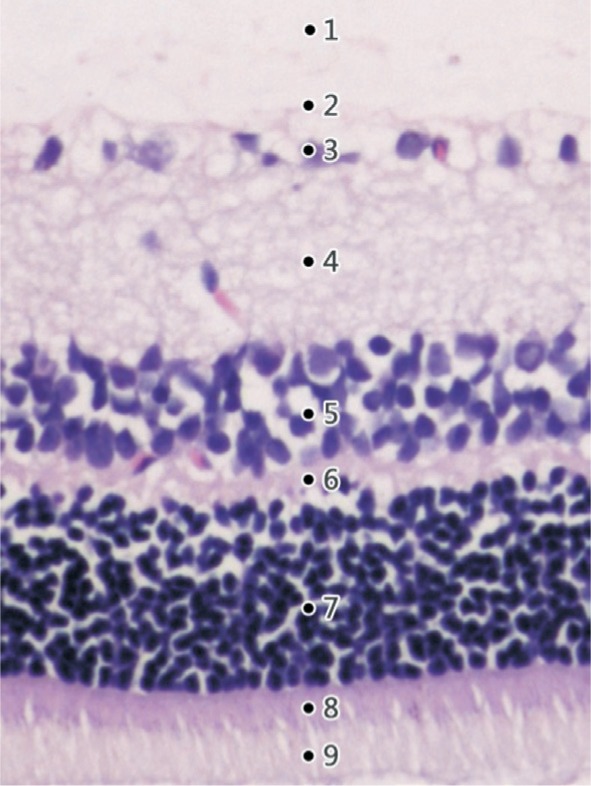
Figure 1. H&E staining of a normal rat's retina (×200) showing well distinguished retinal layers.

1: The inner limiting membrane (ILM); 2: The nerve fiber layer (NFL); 3: The ganglion cell layer (GCL); 4: The inner plexiform layer (IPL); 5: The inner nuclear layer (INL); 6: The outer plexiform layer (OPL); 7: The outer nuclear layer (ONL); 8: The external limiting membrane (ELM); 9: The photoreceptor layer (PL).
