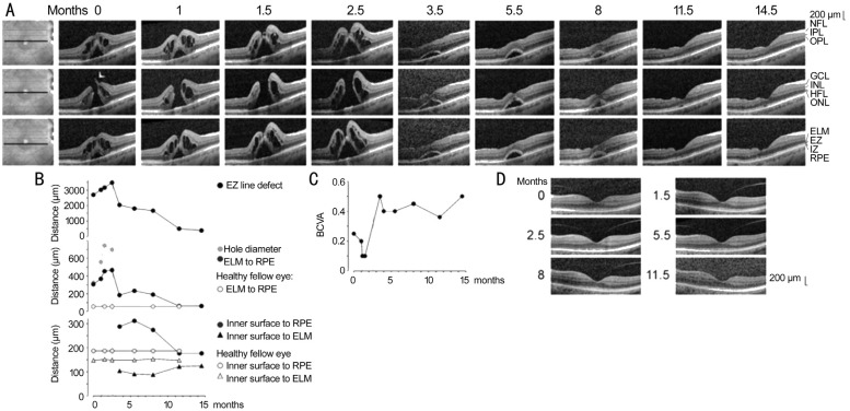
Figure 1. Closure of a macular hole after revitrectomy with autologous platelet concentrate in the right eye of patient 1.
A: Linear horizontal scans through the fovea and parafovea of the right eye. The orientations of the scans are shown at the left side. The months after the examination (0) are indicated above the images. Standard vitrectomy with ILM peeling and revitrectomy with ILM peeling and instillation of autologous platelets were performed 1.1 and 2.6mo after the first visit, respectively. B: Time-dependent alterations of the following parameters measured in linear horizontal scans through the fovea of the right eye: the horizontal diameter of the EZ line defect, the minimum diameter of the macular hole, the vertical distance between the central ELM and the RPE, the vertical distance between the inner surface of the central fovea and the RPE, and the vertical distance between the inner surface of the central fovea and the ELM. In addition, the distances in the fovea of the healthy left eye are shown. The broken vertical lines indicate the time of vitrectomy with ILM peeling. The unbroken vertical lines indicate the time of revitrectomy with ILM peeling and instillation of the platelet concentrate. C: Time dependence of the BCVA. D: Linear horizontal scans through the fovea of the left eye.

