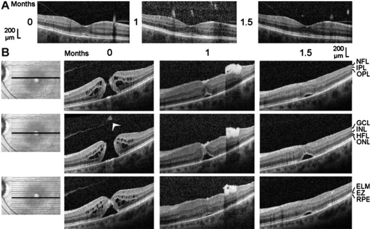
Figure 2. Closure of a macular hole after vitrectomy with autologous platelet concentrate in the left eye of patient 2.
The optical coherence tomographical images were recorded during the first visit (0mo) and 1 and 1.5mo later. A: Linear horizontal scans through the fovea of the right eye. The fovea showed no structural abnormalities with the exceptions of the vitreomacular adhesions at the parafoveas and the thickened hyperreflectivity in the inner layer of the foveola. B: Linear horizontal scans through the fovea and parafovea of the left eye. The orientation of the scans are shown at the left side. Surgery was performed 0.5mo after the first visit. Arrowhead: Operculum.

