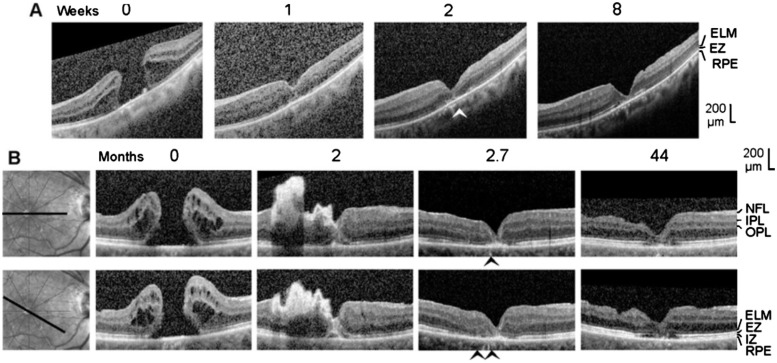
Figure 6. Closure of macular holes after vitrectomy with autologous platelet concentrate in the left eye of patient 6 (A) and the right eye of patient 7 (B).
The images show linear scans through the fovea and parafovea; the orientations of the scans are shown at the left side (B). The weeks (A) and months (B), respectively, after the first clinical examination (0) are indicated above the images. A: Surgery was performed one day after the first visit. B: Revitrectomy with autologous platelet concentrate combined with cataract surgery was performed 1.8mo after the first visit. The arrowheads indicate sites of a direct contact between the central ONL and the RPE.

