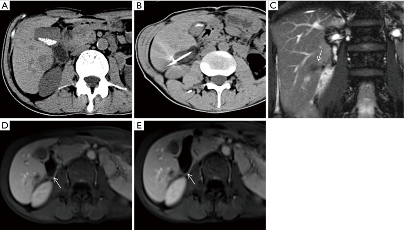
Figure 3.
A 56-year-old male with HCC. (A) CT image revealed a low-density tumor adjacent to the intestine; (B) the tumor was completely covered by ice ball under CT guidance during CA; (C,D,E) follow-up MR scans performed at 1 and 6 months. The tumor presented as hypointensity on coronal T2 imaging (C, arrow). Local tumor recurrence and the damage of the intestine were not found on MR enhancement (D, E, arrows). HCC, hepatocellular carcinoma; CT, computed tomography; CA, cryoablation; MR, magnetic resonance.

