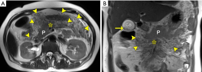
Figure 3.
Retroperitoneal and mesenterical fat necrosis in a 47-year-old woman after an episode of acute necrotizing pancreatitis (extrapancreatic fat necrosis alone). Axial (A) and coronary (B) T2-weighted images without fat-suppression reveal the mesenteric irregular thickening and heterogeneously granular hypointensity (arrowheads) along the root of mesentery (asterisk), indicating the presence of mesenteric fat necrosis. Note the hypointense gallstones (arrow). P, pancreas; GB, gallbladder.

