Fig. 4.

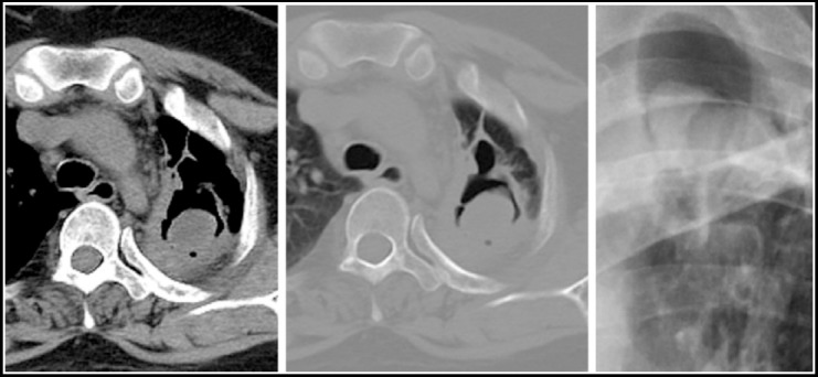
Chest X ray shows a cystic lesion with internal soft tissue opacity and air crescent sign. Corresponding CT chest sections in mediastinal and lung window settings shows a thin walled cavity with a fungal ball.

Chest X ray shows a cystic lesion with internal soft tissue opacity and air crescent sign. Corresponding CT chest sections in mediastinal and lung window settings shows a thin walled cavity with a fungal ball.