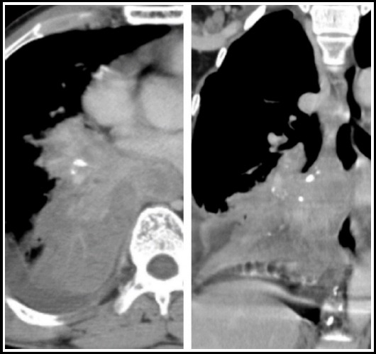
Fig.5.

CT scan chest in mediastinal window settings shows an abnormally enhancing, spiculated infraright hilar mass lesion with specks of calcification with adjacent collapse. This patient received ATT 5 years back and now has biopsy proven bronchogenic carcinoma.
