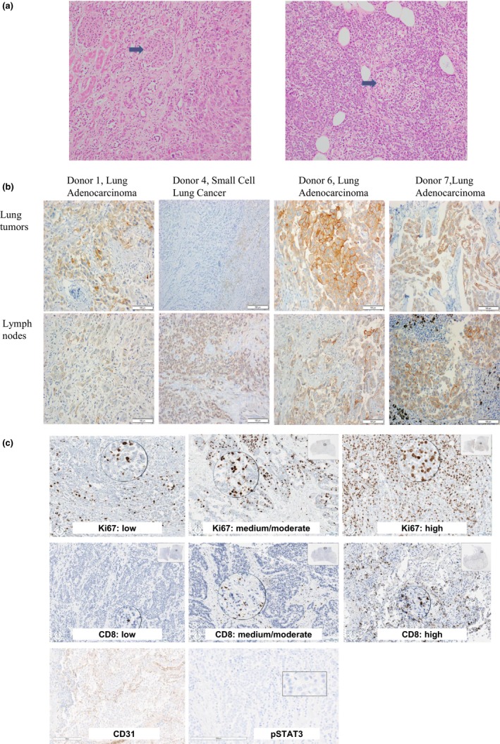
Figure 2.

A, Hematoxylin and eosin stained slides at 200× magnification depicting well‐preserved kidney (on left, arrow indicates preserved glomerulus) and pancreas (on right, arrow indicates preserved pancreatic islet) from rapid tissue donation patient 1. B, Images of slides stained by immunohistochemistry with the E1L3N® anti‐PD‐L1 rabbit monoclonal antibody from one representative lung tumor and one lymph node with metastatic lung cancer from donors 1, 4, 6, and 7 which had tumor with any positivity for PD‐L1 expression. C, Representative images of slides stained by immunohistochemistry with for Ki67, CD8, CD31, and pSTAT3
