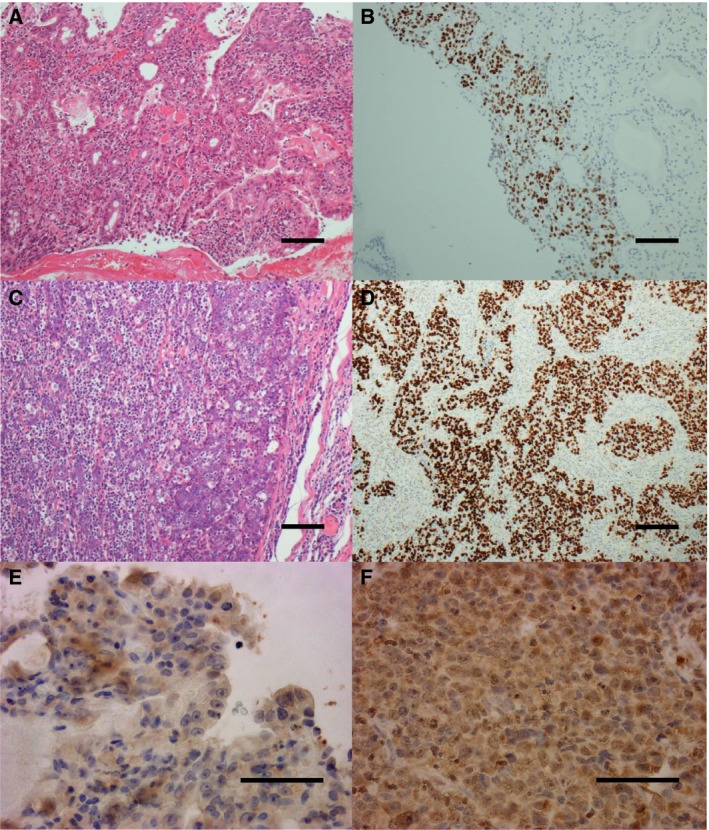
Figure 1.

Histopathological examination of the biopsy from primary and metastatic lesion. (A, C) A metastatic lesion of lymph node and primary lesion were identified as a poorly to moderately differentiated adenocarcinoma (scale bar 100 μm); (B, D) EBER‐in situ hybridization of both lesions showed positive staining (scale bar 100 μm); (E, F) PD‐L1 staining was also positive in both lesions (scale bar 50 μm).
