
Figure 3.
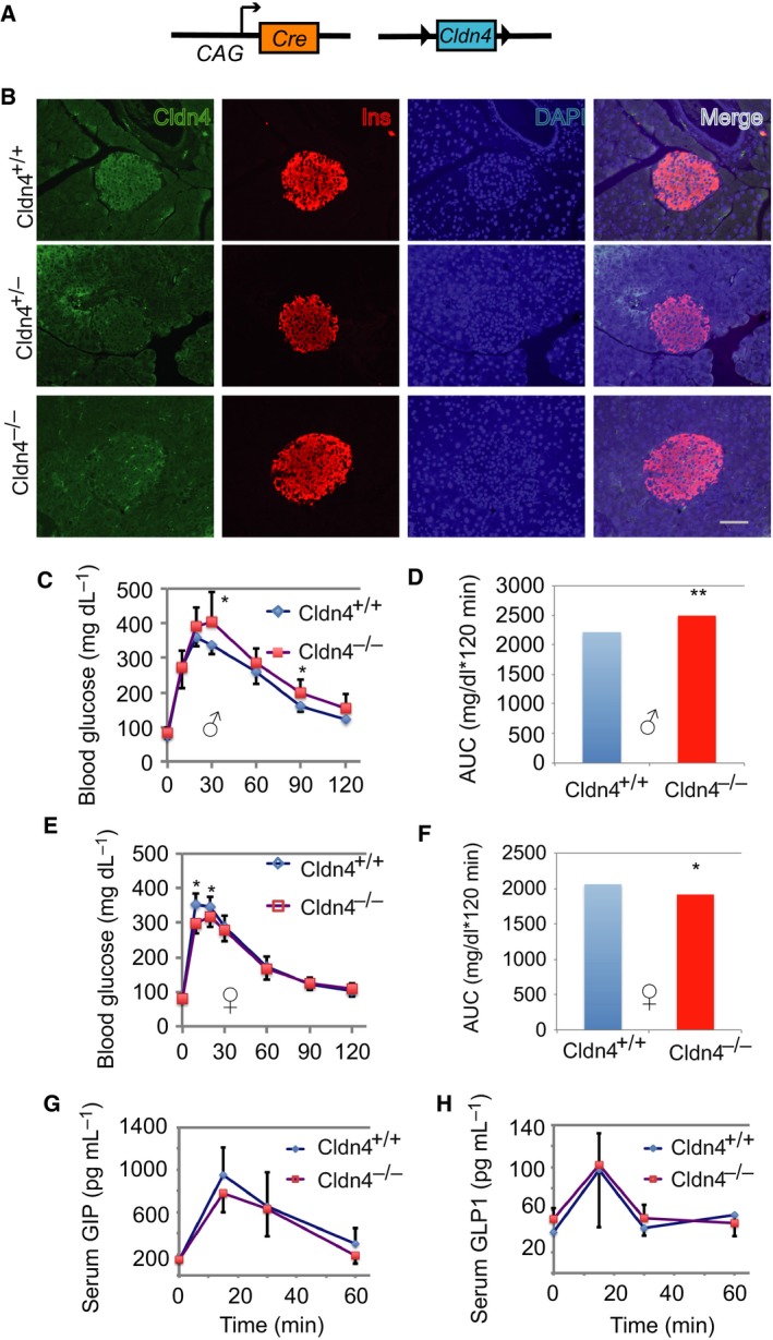
Cldn4 deletion compromises glucose tolerance. (A) Schema showing the Cldn4 deletion strategy. The chicken β‐actin promoter/enhancer coupled with the cytomegalovirus immediate‐early enhancer (CAG) driving Cre‐mediated deletion of the floxed Cldn4. (B) Immunofluorescence analysis. Pancreas sections from Cldn4+/+, Cldn4+/− and Cldn4−/− mice were stained with anti‐Cldn4 (green) and anti‐insulin (Ins, red), and the DNA dye DAPI (blue). Microphotographs were taken under a microscope. Scale bar = 50 μm. (C) Oral glucose tolerance test (OGTT) in males. Cldn4+/+ (n = 9) and Cldn4−/− (n = 8) adult mice were examined. Blood glucose concentrations were determined in the tail vein using an OneTouch UltraVue glucose metre. (D) AUC analysis of OGTT in (C). (E) OGTT in females. Cldn4+/+ (n = 12) and Cldn4−/− (n = 13) adult mice were examined. Blood glucose concentrations were determined as in (C). (F) AUC analysis of OGTT in (E). (F) Serum glucose‐dependent GIP concentrations. Adult male Cldn4+/+ (n = 4) and Cldn4−/− (n = 4) mice were used. (G) GIP concentrations were determined with Bio‐Plex assays. (H) Serum glucagon‐like polypeptide‐1 (GLP1) concentrations. Adult male Cldn4+/+ (n = 3) and Cldn4−/− (n = 3) mice were tested. GLP1 concentrations were determined with Bio‐Plex assays. (C–H) Data presented as mean ± SD, *P < 0.05 and **P < 0.001 compared to Cldn4+/+ (Student's t tests).