
Figure 1.
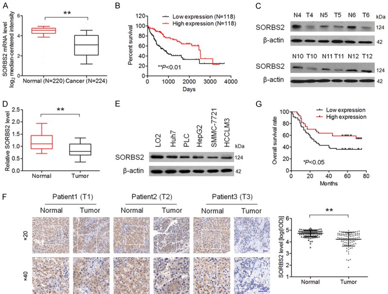
SORBS2 expression is related to HCC progression. A. Analysis based on the oncomine database indicates that SORBS2 mRNA expression was significantly lower in HCC tissue samples compared with normal tissue samples. Data were pooled from two published studies on HCC gene expression [15,16]. B. Relationship between SORBS2 mRNA expression and the overall survival rates of patients with HCC in the TCGA database. C. SORBS2 protein expression was examined in 12 paired tumor (T) samples and normal (N) tissues. D. qRT-PCR analysis of SORBS2 mRNA expression in 12 paired tumor (T) and normal (N) tissues. E. Western blotting analysis of SORBS2 expression in five HCC cell lines and LO2 cells. F. IHC staining of clinical tissue samples with an anti-SORBS2 antibody (samples from 102 patients with HCC). Representative clinical samples of HCC stages I, II, and III are presented (Left). Scale bar, 50 μm. Quantitative analysis of SORBS2 staining revealed a significantly lower staining intensity in HCC samples compared with normal tissue samples (Right). The integrated optical density (IOD) at the same level (× 20) from three sections per sample was measured using Image-ProPlus 6.0 software. G. Low intensity of SORBS2 immunostaining was strongly associated with poor survival rates of patients with HCC (n = 44 in the SORBS2 high-expression group, n = 58 in the SORBS2 low-expression group). **P < 0.01.