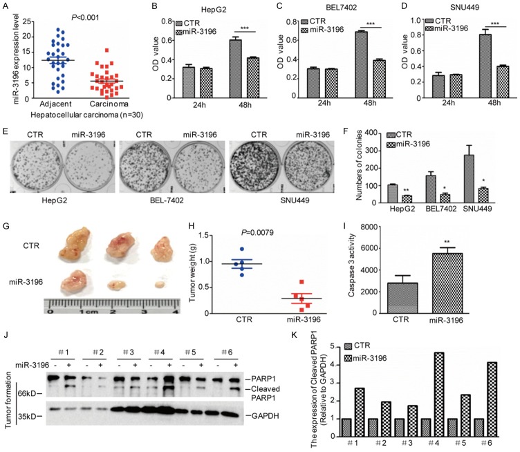
Figure 1.
miR-3196 inhibits HCC cell growth. (A) The expression levels of miR-3196 were analyzed by q-RT-PCR in HCC tissues (n=30) and the adjacent tissues (n=30). (B-D) miR-3196 was transfected into HepG2, SNU449 and BEL7402 cells and the cell proliferation was examined by MTS assay. Data represent the mean ± SD of three independent experiments. ***P<0.001 vs. control. (E, F) The cell growth was examined by a colony formation assay. Data represent the mean ± SD of three independent experiments. **P<0.01 and ***P<0.001 vs. control. (G-K) The tumour-forming abilities of SNU449 cells with or without miR-3196 overexpression were measured in vivo (G). The tumor weight was assessed (H). The Caspase 3 activity was examined (I) and cleaved PARP1 were analyzed by western blotting (J and K).

