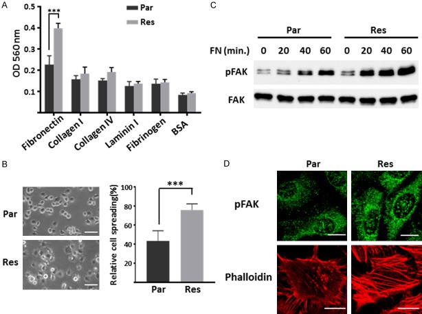
Figure 2.
Detecting the FN-induced cell adhesion, FAK signaling and actin filament formation in Par and Res cells. A. Adhesion ability of Eca109-Par and Res cells upon various ECM proteins. Cell suspensions were planted on the ECM-coated plate for 40 min at 37°C. Attached cells were stained and checked by colorimetric detection. The quantitative data were presented as the means ± standard derivation from three independent experiments (***, P < 0.001 by two-tail unpaired t-test). B. Eca109-Par and Res cells were detached, suspended in serum-free RPMI 1640 for 1 h, and then plated on the FN-precoated (10 µg/ml) dishes. After incubation for 40 min, spread cells were fixed with PFA, and the representative photos were taken (left panel; Scale bar, 50 µm). The percentages of adherent cells were statistically analyzed as the means ± standard derivation of three independent experiments (***, P < 0.001 by two-tail unpaired t-test). C. Eca109-Par and Res cells were plated on the FN-precoated (10 µg/ml) dishes for indicated times. Cell lysates were collected and then immunoblotted by anti-pFAK and anti-FAK antibodies. D. Immunofluorescence staining of p-FAK (top) and phalloidin (bottom) in Eca109-Par and Res cells. Cells were cultured on FN-coated coverslips, then cells were fixed, permeabilized, and visualized with p-FAK and phalloidin-Alexa Fluor 549 (actin), respectively. The images were taken by confocal microscopy. Scale bar, 20 µm.

