
Fig. 5.
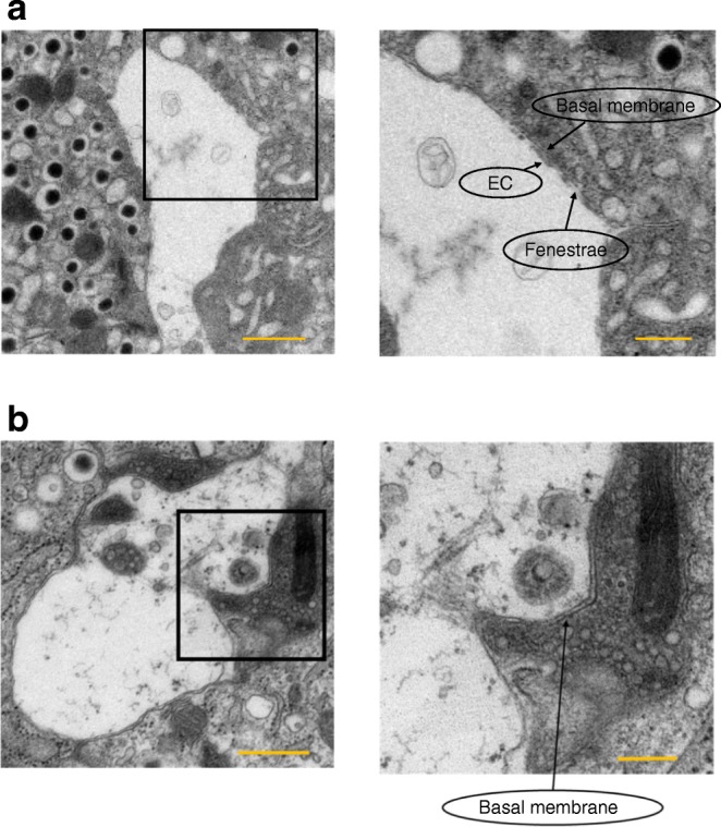
Vascular structure of islets from VE-PDPK1-KO mice and control flox mice. Electron microscopic images showing capillaries surrounded by pancreatic beta cells in (a) control flox mice and (b) VE-PDPK1-KO mice. EC, endothelial cells. Images are representative of four mice. Magnification × 5000 (a, left) or × 3000 (b, left) scale bars, 1 μm; or × 10,000 (right), scale bars, 500 nm