
Figure 3.
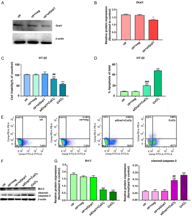
Effects of siOrai1 knockdown on cell proliferation and apoptosis. A, B. After transfection of HT-22 cells with siOrai1 for 72 h, the extracted proteins were subjected to western blotting with the Orai1 antibody. β-actin was used as a control for equal protein loading. Data represent the means ± SD from three independent experiments. C. After transfection of HT-22 cells with siOrai1 for 6 h, cells were then incubated with 100 µM CoCl2 for 24 h. Cell viability was assessed by an MTT assay, and the absorbance values of the treated samples are expressed as percentages of the absorbance values of the control samples. D, E. After transfecting HT-22 cells with siOrai1 for 6 h, cells were incubated with 100 µM CoCl2 for 24 h and then stained with Annexin V-FITC/PI for analysis of cell apoptosis by flow cytometry. F, G. After transfection of HT-22 cells with siOrai1 for 6 h, cells were incubated with 100 µM CoCl2 for 72 h, and the extracted proteins were subjected to western blotting with Bcl-2 and cleaved-caspase-3 antibodies. β-actin was used as a control for equal protein loading. Data are expressed as means ± SD from three independent experiments. **P<0.01 and ***P<0.001 vs. Control; #P<0.05, ##P<0.01, and ###P<0.001 vs. CoCl2 (ctr, untreated cells; ctr+neg, untreated cells incubated with siOrai1 negative control buffer; ctr+siOrai1, untreated cells incubated with siOrai1 buffer; siOrai1+CoCl2, cells were transfected and then treated with CoCl2; CoCl2, cells were treated with CoCl2).