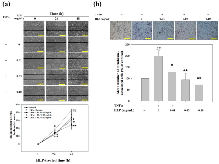
Figure 4.
Effect of HLP on TNF-α-induced A7r5 cell motility and invasion. (a) Monolayers of A7r5 cells treated with TNF-α (10 ng/mL) in the absence or presence of various concentrations (0, 0.01, 0.05 and 0.10 mg/mL) of HLP were scraped and the number of cells in the denuded zone was photographed and quantified after indicated times (0, 24, and 48 h). Quantitative assessment of the mean number of cells in the denuded zone was presented as mean ± SD (n = 3) from three independent experiments. (b) A7r5 cells were treated with TNF-α in the absence or presence of various concentrations of HLP for 24 h. Invasion assay was performed using Boyden chamber. Representative photomicrographs of the membrane-associated cells were assayed by Giemsa stain. The purple parts indicate the membrane-associated cells. “% of control” denotes the mean number of cells in the membrane expressed as a proportion of that control group. Images were taken at 200× magnification; scale bar, 30 μm. The quantitative data are presented as mean ± SD (n = 3) from three independent experiments. # p < 0.05, ## p < 0.01 compared with the control. * p < 0.05, ** p < 0.01 compared with the TNF-α group. +, added; −, non-added.

