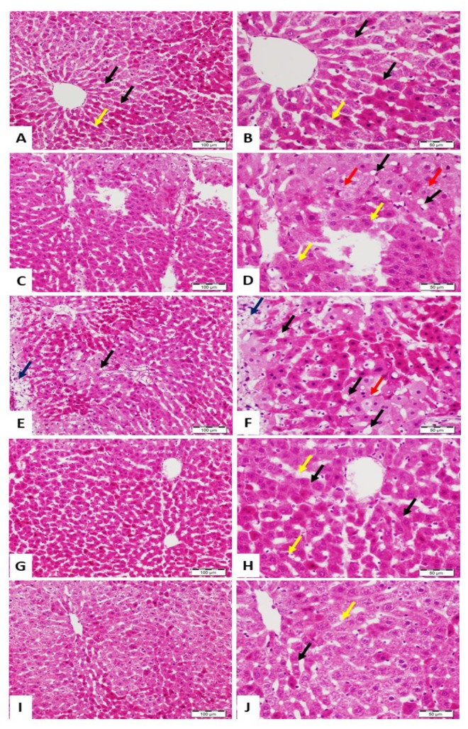
Figure 2.
Photomicrographs of hematoxylin and eosin (H&E)-stained sections from liver of (A,B) control rats showing normal structure and architecture with hepatocytes arranged in thin plates (black arrow), sinusoids (yellow arrow), and central vein; (C–F) Pb(II)-intoxicated rats showing distorted lobular architecture, ballooning (black arrow), multinucleated hepatocytes (yellow arrow), microsteatotic changes (red arrow), and large areas with necrosis (blue arrow); (G,H) Pb(II)-administered rats treated with ALRE showing normal hepatic tissue with normal hepatocytes (black arrow) and sinusoids (yellow arrow); and (I,J) Pb(II)-administered rats treated with Vit. C normal hepatocytes (black arrow) and sinusoids (yellow arrow). (A, C, E, G, and H: ×200, Scale bar 100 µm) and (B, D, F, H, and J: ×400, Scale bar 50 µm).

