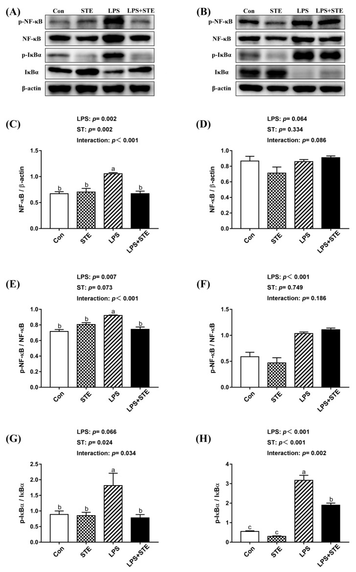
Figure 2.
Effects of stevioside supplementation on the protein expression of NF-κB, p-NF-κB, IκBα and p-IκBα. (A) Western blot analysis of NF-κB, p-NF-κB, IκBα and p-IκBα in the jejunal mucosae. (B) Western blot analysis of NF-κB, p-NF-κB, IκBα and p-IκBα in the ileal mucosae. (C) Statistical analysis of NF-κB/ β-actin in the jejunal mucosae. (D) Statistical analysis of NF-κB/ β-actin in the ileal mucosae. (E) Statistical analysis of p-NF-κB/ NF-κB in the jejunal mucosae. (F) Statistical analysis of p-NF-κB/ NF-κB in the ileal mucosae. (G) Statistical analysis of p-IκBα/ IκBα in the jejunal mucosae. (H) Statistical analysis of p-IκBα/ IκBα in the ileal mucosae. CON, non-challenged broilers fed a basal diet; STE, non-challenged broilers fed a basal diet supplemented with 250 mg/kg stevioside; LPS, LPS-challenged broilers fed a basal diet; LPS + STE, LPS-challenged broilers fed a basal diet supplemented with 250 mg/kg stevioside. Data are presented as mean value ± SEM (n = 6).a,b,c Means with different letters are significantly different (p < 0.05).

