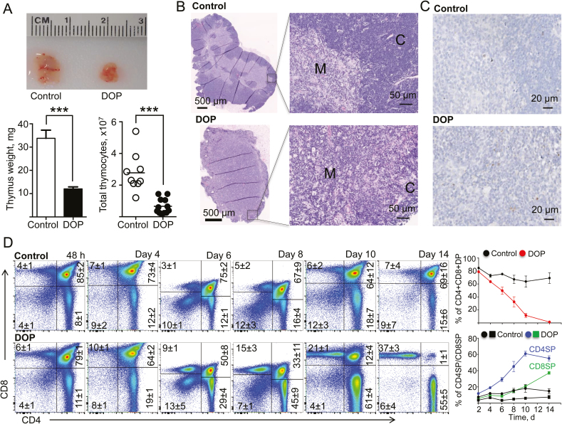
FIGURE 7.
S1P lyase inhibition promoted atrophy of the thymic cortex and depletion of stage II (double-positive CD4/CD8) T cells in TNFΔARE mice. A, Representative images, weights (mg), and total number of thymocytes from TNFΔARE mice treated with DOP or vehicle control for 2 weeks. B, Representative hematoxylin and eosin–stained sections and (C) cleaved-caspase-3 staining of the thymus of DOP- and vehicle-treated TNFΔARE mice for 2 weeks (C, cortex; M, medulla). D, Representative dot plots and CD4/CD8 percentages of thymocytes from TNFΔARE mice treated with DOP or vehicle for 2 weeks or indicated times. Data are expressed as mean ± SEM; n ≥ 5 mice/group/time point (***P < 0.001 by 2-tailed t test). Abbreviations: DP, double-positive (CD4+/CD8+) cells; SP, single-positive mature CD4+CD8- or CD4-CD8+ cells.

