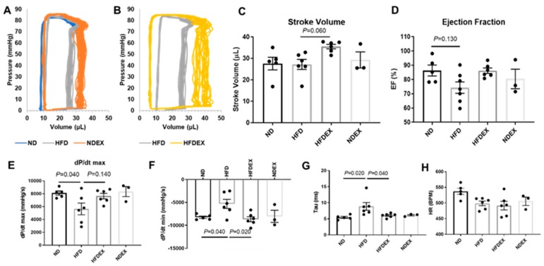
Figure 3.
Exercise training improves diastolic left ventricle dysfunction in diet- induced obese mice. (A) Representative PV trace in ND, HFD, and NDEX mice. (B) Representative PV trace in HFD mice (trace replicated from A) and HFDEX mice. (C) Stroke volume was elevated in HFDEX mice compared to HFD. (D) Ejection fraction (EF) was not changed with HFD or EX. (E) Maximum change in pressure over time (dP/dtmax) was reduced with HFD. (F and G) Measures of diastolic function (dP/dtmin and Tau) were worsened with HFD but normalized in HFDEX mice. (H) Heart rate (HR) during the recordings was constant between groups. All values are expressed as mean ± SEM with dots representing each animal. One-way ANOVA and Tukey’s post-hoc test were used for statistical analysis.

