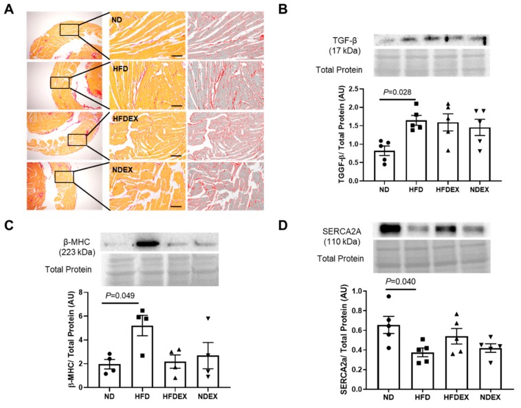
Figure 4.
High- fat diet upregulates markers of cardiac structural remodeling. (A) Representative sirius red staining of a section of the left ventricle for interstitial collagen fibers was unaltered in all the four groups. Scale bar = 200 µm. A similar portion of the left ventricular wall was imaged in all groups. (B) Western blot quantification of the pro-fibrotic signaling marker TGF-β protein expression in left ventricle tissue demonstrates an upregulation in HFD mice compared to ND. (C) Western blot quantification of the hypertrophy marker β-MHC protein expression in left ventricle tissue demonstrates an upregulation in HFD mice compared to ND. (D) Western blot quantification of contractile protein sarco/endoplasmic reticulum Ca2+-ATPase (SERCA-2A) protein expression in left ventricle tissue demonstrates a downregulation in HFD mice compared to ND. All values are expressed as mean ± SEM with dots of different shape representing each animal in a group. One-way ANOVA and Tukey’s post-hoc test were used for statistical analysis.

