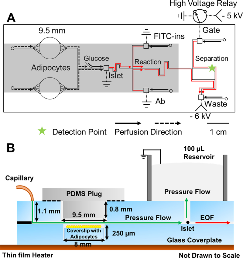
Figure 1.

Microfluidic chip layout. (A) Top view of chip illustrating the channel layout, electrical connections, and flow directions for co-culture and monitoring insulin. Solid lines indicate the 15 μm deep and 50 μm wide microfluidic channels. Dotted lines indicate electrical connections. The 9.5 mm circles indicate the adipocyte chambers drilled through the glass. Smaller circles indicate perfusion inlets, and squares indicate reservoirs that are sampled by electroosmotic flow. Electroosmotic flow directions are indicated by the red arrows. All fluidic inlets are 360 µm diameter. Reaction refers to the portion of channel where immunoassay reagents mix, and reaction happens. Arrows indicate perfusion flow direction. Dashed arrow lines indicate direction of flow during 1st step of experiment of perfusion co-culture for 3 h. Islets are co-cultured with adipocytes for 3 h without chemical monitoring. Solid arrow lines indicate flow during 2nd step of experiment of glucose stimulation. Perfusion from adipocytes (dashed lines) is stopped and glucose is delivered from the side channel to stimulate insulin secretion from the islets. Step changes in glucose concentration of 3–11 mM are made using external syringe pumps and valves. The relay is switched to inject sample onto the electrophoresis channel every 8 s. The shaded portion of the chip indicate parts that are heated during experiments with a thin film resistive heater. Laser-induced fluorescence (LIF) detection point occurred 1 cm past injection cross, as indicated by the star. -HV is where high voltage is applied. (B) Side view of adipocyte and islet perfusion culture. Adipocytes and islet are loaded into cell chambers and perfused with pressure-driven flow from the capillary. Perfusate flows into the islet chamber and up into a 100 μL fluidic reservoir. The adipocyte chambers are sealed with a PDMS plug. Solution with insulin from the chamber is sampled by electroosmotic flow (EOF in the drawing) through the sampling channel.
