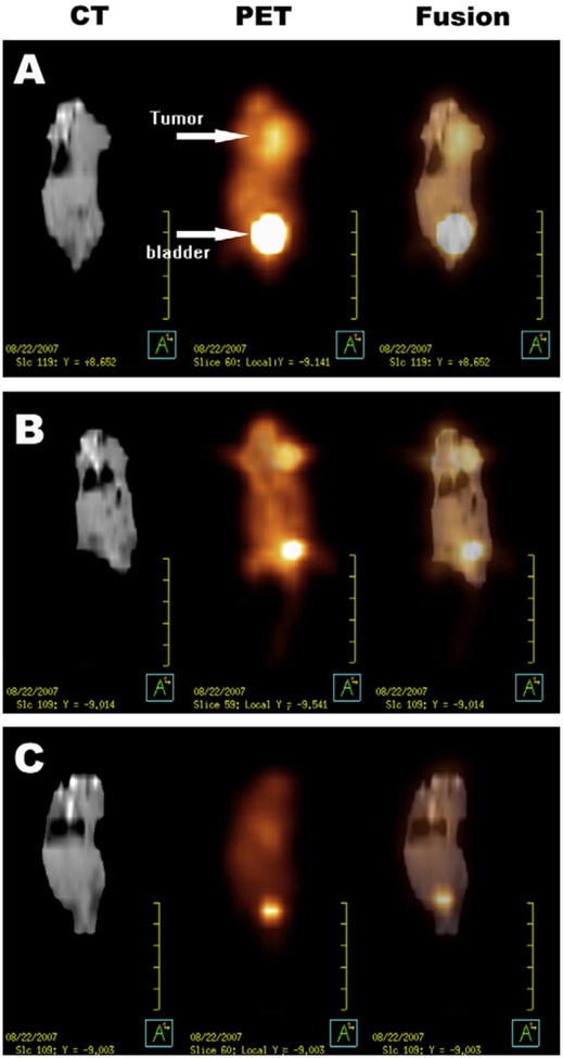
Figure 3. Male ICR mice bearing a subcutaneous H22 (murine hepatoma cell line) tumor at the left side of the thorax.
A) Coronal images of a mouse in the control group (saline). B) Coronal images of a mouse in the group receiving intratumoral free cisplatin (5 mg/kg). C) Coronal images of a mouse in the group receiving intratumoral cisplatin-loaded nanoparticles (5 mg/kg). Reproduced from Li et al.28

