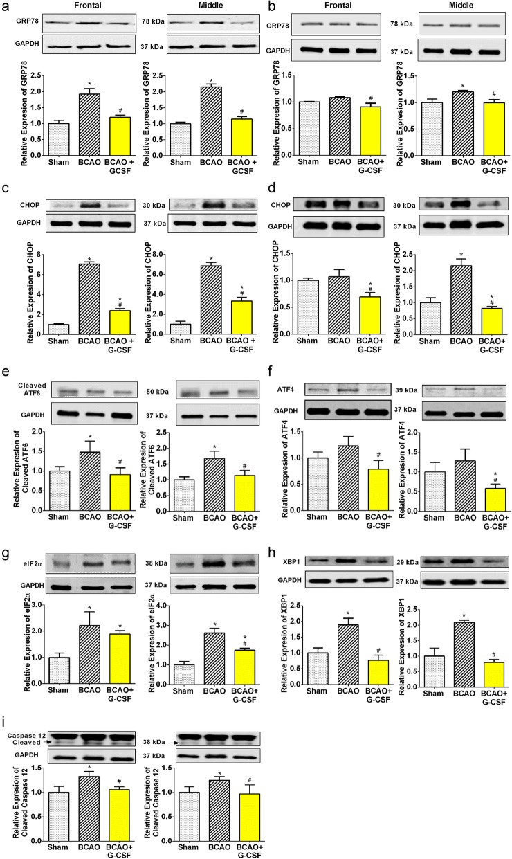
Fig. 2.
Effect of G-CSF on expression of GRP78, CHOP and ER stress proteins. a GRP78 levels in the frontal (F (2, 18) = 9.549, p = 0.0015) and middle F (2, 12) = 283.3, p < 0.0001) region of brain on day 4. b GRP78 levels in the frontal (F (2, 15) = 4.378, p = 0.0318) and middle (F (2, 14) = 4.787, p = 0.0261) region of brain on day 7. c CHOP levels in the ischemic frontal (F (2, 21) = 10.77, p = 0.0006) and middle (F (2, 11) = 74.31, p < 0.0001) region of brain on day 4. d Levels of CHOP in the ischemic frontal (F (2, 14) = 6.101, p = 0.0473) and middle (F (2, 20) = 26.63, p < 0.0001) region of brain on day 7. e Cleaved ATF6 levels in the frontal (F (2, 27) = 5.464, p = 0.0102) and middle (F (2, 26) = 6.514, p = 0.0051) region of brain. f ATF4 levels in the frontal (F (2, 15) = 4.084, p = 0.0384) and middle (F (2, 13) = 3.965, p = 0.0452) region of brain. g eIF2α levels in the ischemic frontal (F (2, 8) = 5.671, p = 0.0293) and middle (F (2, 11) = 21.24, p = 0.0002) regions of brain. h XBP1 levels in the frontal (F (2, 12) = 10.29, p = 0.0025) and middle (F (2, 7) = 19.53, p = 0.0014) region of brain. i Cleaved caspase-12 levels in the ischemic frontal (F (2, 9) = 8.165, p = 0.0095) and middle (F (2, 11) = 5.357, p = 0.0238) regions of brain. Representative western blots are presented with cropped blot panels showing target protein signals and control (GAPDH) protein signals in separate panels derived from the same gel. Graphs shows mean ± SEM. * and # significant compared to sham & vehicle treated groups, respectively by ANOVA and Tukey post hoc tests (n = 5, p < 0.05)

