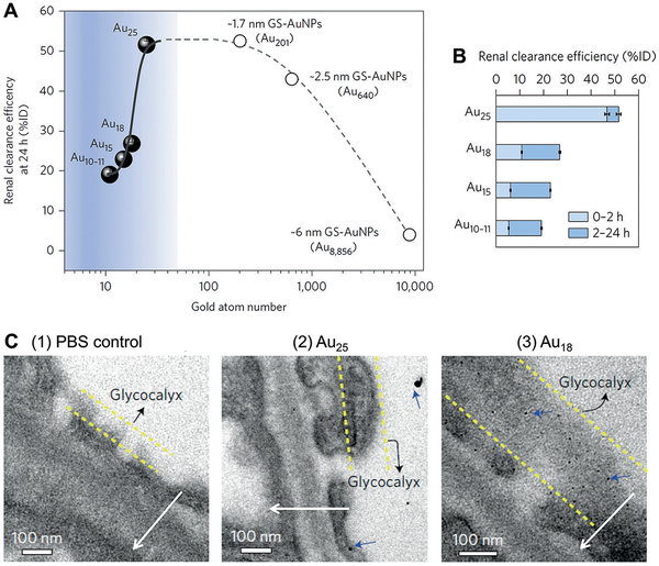
Figure 3.
Particle size effect on the renal clearance of glutathione-coated gold nanoparticles (GS-AuNPs) from 6 nm to sub-nanometer regime. A) Renal clearance efficiencies of Au10–11, Au15, Au18, and Au25, and GS-AuNPs with core sizes of 1.7 nm (Au201), 2.5 nm (Au640), and 6 nm (Au8856) at 24 h after intravenous injection vs. number of gold atoms. Percentage of injected dose, %ID. B) Renal clearance efficiencies of Au10–11, Au15, Au18, and Au25 at 0–2 h and 2–24 h after intravenous injection (n =3 for Au10–11, Au15, and Au25; n= 6 for Au18). C) Electron microscopy (EM) images of the ultrastructure of the glomerular filtration membrane from mice at 10 min after intravenous injection of phosphate buffered saline (PBS control), Au25 or Au18. Although gold clusters were too small to be directly revealed with EM, they can be visualized with EM after silver staining that increases the particle core size and contrast. (1) No silver nanoparticles were observed in the control glomeruli after silver staining and (2) only a few large silver-enhanced Au25 were found in the glomeruli of the mice injected with Au25. (3) In contrast, a large number of monodispersed silver-enhanced Au18 (blue arrows) were retained on the glycocalyx of the endothelium and podocytes. Arrows point the direction of filtration from vascular lumen to Bowman’s space. Reproduced with permission from Ref. [47]. Copyright 2017, Nature Publishing Group.

