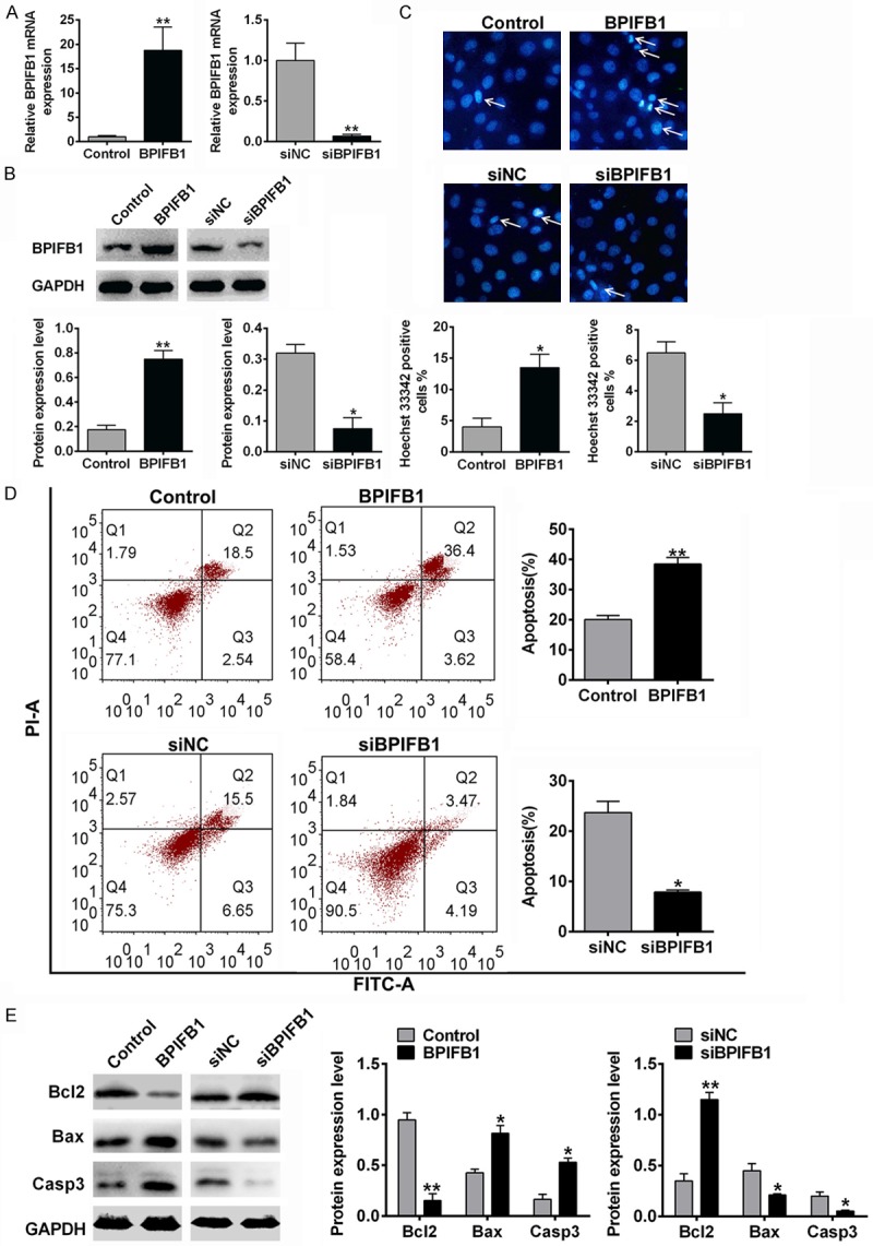
Figure 1.

The effects of BPIFB1 on the cell apoptosis of NPC-KT cells. NPC-KT cells were transfected with BPIFB1 mimics or siRNA and the negative control (A, B) BPIFB1 expression of mimics or siRNA transfected NPC-KT cells was measured by PCR (A) and Western blotting (B). (C) DNA damage was measured by Hoechst staining. (D) The cell apoptosis was analyzed by flow cytometry. (E) The apoptosis related protein expression was assessed by Western blotting. Data were presented as the mean ± SD. n=3, *P<0.05, **P<0.01 vs. Control.
