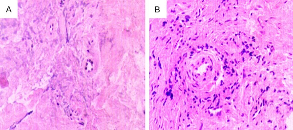
Figure 1.

Histological observations of tissues both from the healthy and periapical granuloma groups using H E staining. A. A small number of infiltrating inflammatory cells were observed in the healthy group specimens. B. Numerous infiltrating inflammatory cells, capillaries, and fibroblasts were observed in the periapical granuloma specimens. (original magnification, ×400).
