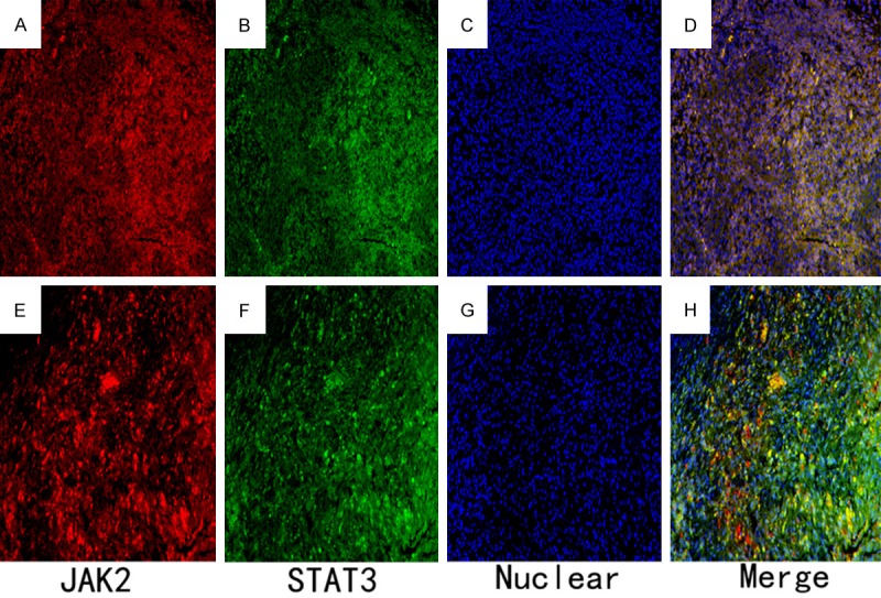
Figure 4.

Double immunofluorescence staining observations for the healthy group and the periapical granuloma group. Immunofluorescence staining for the JAK2- and STAT3-positive cells was conducted followed by Alexa Fluor 555 labeling of donkey against rabbit lgG (H+L) (red) and Alexa Fluor 488-labeled goat anti mouse IgG (H+L) (green). Nuclear counterstaining was performed using 40,6-diamidino-2-phenylindole (blue). A. Slightly JAK2-positive cells were observed in the healthy control group. B. Minimal intense immunoreactions for STAT3-positive cells were also found in healthy control group. C. Nuclears were stained for the healthy control group. D. The merged image indicates that some JAK2-positive cells are positive for STAT3. E. Intense immunoreactions for JAK2-positive cells were observed in periapical granuloma group. F. Numerous STAT3-positive cells were also found in periapical granuloma group. G. Nuclears were stained for granuloma group. H. The merged image suggests that most JAK2-positive cells are also positive for STAT3 (×200).
