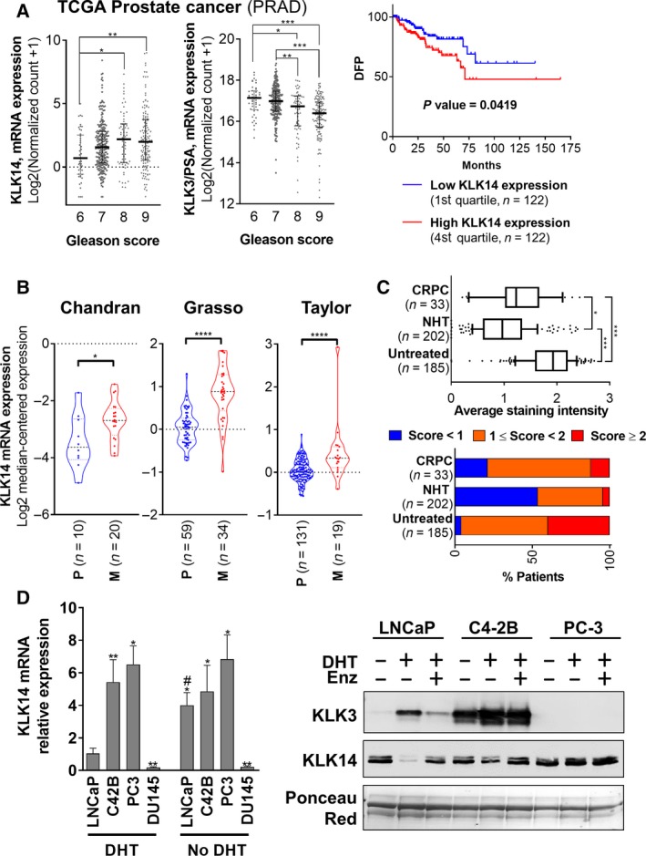
Figure 1.

KLK14 expression is associated with advanced PCa. (A) Expression of KLK14 and KLK3/PSA mRNA in the TCGA prostate adenocarcinoma dataset. Left: Expression of KLK14 mRNA increases with Gleason score of PCa. KLK3/PSA mRNA is shown for comparison. Results are presented as median ± interquartile range. Right: Patients with high levels of KLK14 mRNA in the primary tumor (4th quartile, 122 samples) have a significant shorter DFP compared to patients with low levels of KLK14 expression in the primary tumor (1st quartile, 122 samples). (B) Expression of KLK14 mRNA in PCa‐derived metastases (Chandran, Grasso, Taylor datasets, Oncomine data portal; median ± interquartile range). KLK14 mRNA is significantly elevated in PCa‐derived metastases (M) compared to primary tumor (P). (C) Top: Average KLK14 expression determined by IHC on primary tumors from 185 untreated patients, 202 patients responsive to neoadjuvant hormonal therapy (NHT), and in 33 patients who developed CRPC. Results are presented as median ± interquartile range and 10–90 percentile. Bottom: Percentage of samples scored with low (< 1), medium (1 ≤ score < 2) and high (score > 2) staining per patient groups. (D) Left: Expression of KLK14 (mRNA level, RTqPCR, mean ± SD) in androgen‐dependent LNCaP cells and androgen‐independent and metastatic PCa cells (C42B, PC‐3 and DU145 cells) grown in CSS ± 10 nm DHT for 48h. N = 3, *P < 0.05, **P < 0.01, ***P < 0.001, compared to LNCaP cells DHT condition, # P < 0.05, compared to the respective DHT condition for each cell line. Two‐way ANOVA test was performed. Right: KLK14 and KLK3/PSA protein expression (western blot) in concentrated conditioned media from LNCaP, C4‐2B, and PC‐3 cells grown in serum‐free medium ± 10 nm DHT ± 10 µm Enz (enzalutamide) for 72 h.
