
Figure 5.
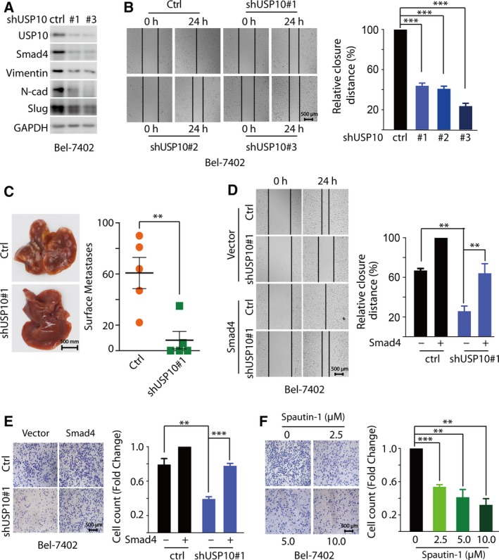
USP10 depletion Inhibits HCC metastasis through down‐regulation of Smad4. (A) Depletion of USP10 in Bel‐7402 cells significantly decreased the expression of EMT marker proteins (N‐cadherin, vimentin, and Slug). Bel‐7402 cells were infected with lentivirus encoding the indicated shRNAs, and cell lysates were immunoblotted with indicated antibody. (B) Depletion of endogenous USP10 with shRNAs markedly decreased cell migration by wounding healing assay. Bel‐7402 cells infected with lentivirus encoding the indicated shRNAs were treated with TGF‐β (5 ng·mL−1) for 24 h. (C) Depletion of USP10 in Bel‐7402 cells significantly decreased liver surface metastatic nodules of BALB/c nude mice. A representative image of liver was shown, and liver surface metastatic nodules were enumerated. (D, E) Overexpression Smad4 reversed cell migration by wounding healing assay (D) or transwell migration assays (E). Bel‐7402 cells infected with lentivirus encoding the indicated shRNAs and (or) plasmids were treated with TGF‐β (5 ng·mL−1) for 24 h. (F) Spautin‐1 inhibited HCC metastasis in a dose‐dependent manner. Bel‐7402 cells were treated with TGF‐β (5 ng·mL−1) and the indicated concentrations of Spautin‐1 (0, 2.5, 5, and 10 μm) for 24 h. The results represent the means (±SD) of three independent experiments. **P < 0.01, ***P < 0.001.