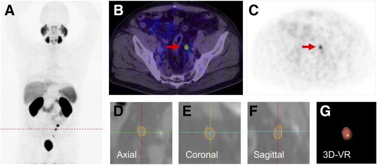
FIGURE 2.
A 58-y-old patient with PSA relapse of 4.9 ng/mL, 9 mo after prostatectomy (GS, 6; iPSA, 10 ng/mL). (A) Patient presented multiple 68Ga-PSMA PET–positive nodes as shown in PET maximum-intensity projection. (B and C) A positive node (red arrow) with PSMA uptake SUVmax 7.2 was segmented using Fraunhofer MEVIS software (SAD, 5.93 mm; LAD, 7.73 mm; and volume, 0.31 mL) Images (D–F) show segmented lymph node on each plane and volume rendering (VR) (G).

