
Figure 1.
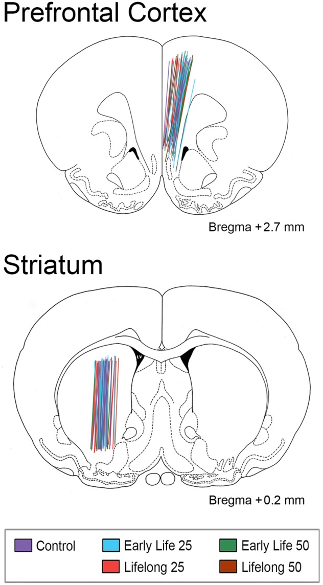
Brain atlas images and coordinates (Paxinos and Watson, 1997) depicting the central axes and active lengths (4 mm) of each microdialysis probe implanted in right medial prefrontal cortex (mPFC) and left striatum (STR) delineated by experimental groups (see legend). It is clear that probe implantation sites across experimental groups were not distinguishable.