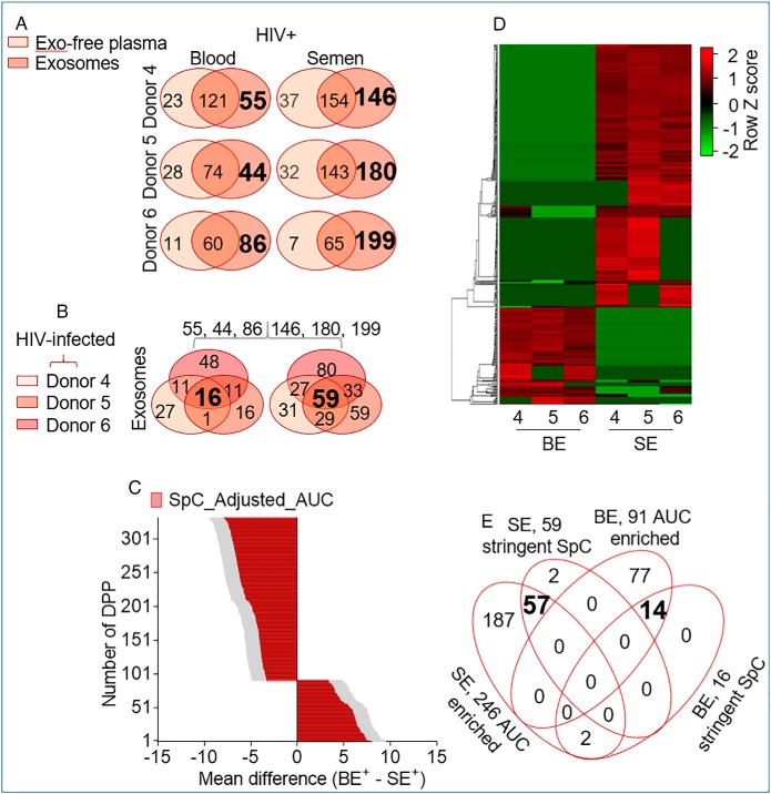
Fig. 6.
Profiling exProteins enriched in blood and semen exosomes from HIV infected participants. A, Two-way Venn diagram of proteins in the exosome and exosome-free compartments showing donor-dependent variation in protein compartmentalization and identifying BE and SE enriched proteins. Exosome-specific proteins are in large and bold font. B, Three-way Venn analysis identifying proteins from that are common to BE and SE in all donor samples. Common proteins for each exosome type is in large and bold font. C, Quantitative intensity profile of the SpC_ adjusted_AUC identified differentially present proteins (DPP) in BE and SE. Statistics was performed by Two-way ANOVA with the Benjamini and Hochberg original FDR method. Adjusted p values are shown in supplemental Table S4. Differences were significant when adjusted p value < 0.05. D, Heatmap of BE and SE DDP revealing body-fluid specific differences with minor donor-dependent variabilities. E, Four-way Venn diagram integrating exosomal proteins from the SpC and the DPP data sets.

